News

Many congratulations to Dr. Udayasankar and his co-authors Dr. Kalb, Dr. Desoky, Dr. Gilbertson & Dr. Martin for being awarded a bronze medal for their outstanding work. The American Roentgen Ray Society has awarded their electronic exhibit abstract, E1823, entitled, Non-Enhanced MRI of the Abdomen in Comprehensive Evaluation of Acute Abdominal Pain in Children: A Paradigm Shift, with an ARRS 2019 Bronze medal for this year’s ARRS Scientific Program.

Dr. Harrison Barrett is retiring. Please join us for a celebration of Harry’s remarkable career and his upcoming 80th birthday! A two-day event is planned for Thursday, May 2nd and Friday, May 3rd, 2019.

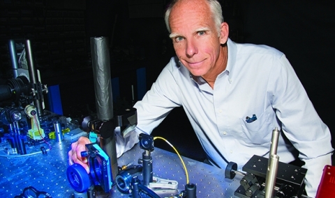
Harrison Barrett, Regents' Professor of medical imaging, optical sciences and applied mathematics, received the distinction last month. and will be inducted in April at the NAI's annual meeting in Houston.

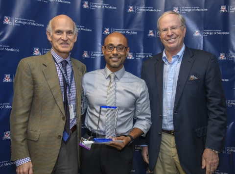
Dr. Bobby Kalb was among the first recipients of the College of Medicine-Tucson’s Clinical Excellence Award. He was honored during the General Faculty Meeting. We are honored to have him on our team.

Dr. Harrison Barrett, Regents Professor of Optical Sciences, Medical Imaging, and Applied Math, was honored at a reception celebrating the establishment of the Harrison H. and Catherine C. Barrett Endowed Chair in Optical Sciences for Cancer Imaging.

Dr. Andrew Karellas, PhD, DABR, FAAPM, FACR, Professor, Medical Imaging, Vice-Chair of Faculty Development, received a certificate of recognition from the College of Medicine Dean’s Council on Faculty Affairs Committee

Zhonglin Liu, PhD, has been awarded an NIH/NCI Clinical and Translational Exploratory/Developmental Studies Grant (R21) of $426,389 for his research project entitled “Novel molecular probes for imaging colorectal cancer.” Dr. Liu will be collaborating with Dr. Brian Gray (Co-PI) at Molecular Targeting Technologies, Inc. (MTTI).
A vetted list of reliable organizations that will give you information on how to help Puerto Rico. This list will be updated every day.
Tom Kenyon, MD, MPH, CEO of HOPE (and a UAHS Pediatrics graduate, 1982) is asking UAHS professionals to help in Puerto Rico. At this time, Project HOPE is actively engaged with emergency response efforts in Puerto Rico. HOPE has a team on the ground to conduct an initial needs assessment to see where Project HOPE can fill a gap, and make an impact. There is a high demand for volunteers with Spanish-speaking skills.
Dr. Marvin “Marv” J. Slepian, Professor of Medicine (Cardiology), Biomedical Engineering and McGuire Scholar in the Eller College of Management at the University of Arizona, has been recently cross-appointed as Professor of Medical Imaging.

Russell Witte, PhD, (Department of Medical Imaging )and Hao Xin, PhD, (Electrical and Computer Engineering Department) have been awarded a $1.44 million grant from the DOD for their research entitled Integrated Platform for Spectroscopic Thermoacoustic Imaging and Focused Microwave Therapy of the Breast.
The Department of Medical Imaging is excited and honored to have two new faculty members - Andrew Karellas, PhD, DABR, FAAPM, FACR, and Srinivasan Vedantham, PhD - to lead a program to develop the next generation of CT scanners for human imaging.

Dr. Lars Furenlid and a team of collaborators have been awarded a grant from the National Institute for Biomedical Imaging and Bioengineering (NIBIB). The project, titled ADAPTISPECT-C: A NEXT-GENERATION, ADAPTIVE BRAIN-IMAGING SPECT SYSTEM FOR DRUG DISCOVERY AND CLINICAL IMAGING, aims at leading the way in understanding the human brain by enabling the development of biomarkers with unprecedented specificity for mapping neuroreceptors and proteinopathies associated with disease and dementia.

Professor Harrison Barrett has received a $2.5 M Grant from the NIBIB to continue the work supported by an R01 that has been continuously funded since 1990 and included a ten-year period NIH MERIT award.

Congratulations to Arthur F. Gmitro, PhD, the 2016 Founders Day lecturer at the UA College of Medicine! Dr. Gmitro will give the 2016 Founders Day lecture at noon, November 17, in Kiewit Auditorium. Founders Day is an occasion when we pause to honor the individuals whose scientific contributions allow the College of Medicine - Tucson to pursue its mission and fulfill its vision. The day is named in recognition of all those people, past and present, who have participated in the establishment and growth of the College.

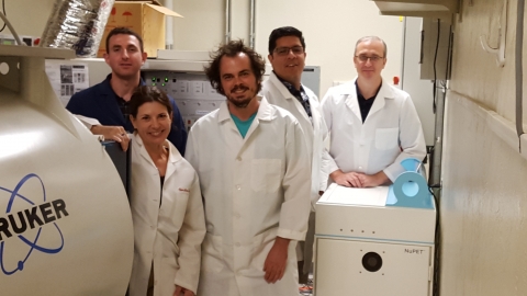
On September 1, 2016, Cubresa Inc., a medical imaging company that develops and markets molecular imaging systems, announced the successful installation of their compact PET scanner called NuPET™ for preclinical PET (Positron Emission Tomography) and MRI (Magnetic Resonance Imaging) in the Department of Medical Imaging at the University of Arizona. The University of Arizona is developing a dual-modality approach by using Cubresa’s NuPET™ PET scanner and a dynamic MRI technique to more fully characterize cancerous tumors.

Mihra Taljanovic MD, PhD, and Lana Gimber, MD, have both earned the prestigious RSNA Honored Educator Award in 2016. This achievement recognizes an individual’s dedication to furthering the profession of radiology and commitment to radiology education by delivering high-quality educational content for RSNA endeavors.

We are pleased to introduce eight new faculty members who have joined the Department of Medical Imaging. All new faculty members will practice at both Banner-University Medical Center Tucson campuses.

Several Department of Medical Imaging doctors have been selected as top doctors by Castle Connolly Ltd. and they are featured in the “Top Doctors” article in the current issue of Tucson Lifestyle (July Issue, page 41). Congratulations to these physicians (below) for being selected as Tucson’s Top Doctors in Tucson Lifestyle!

It’s estimated that approximately 50 percent of all women of reproductive age have uterine fibroids, although not all are diagnosed. Uterine fibroids, also known as leiomyomas or fibromas, are tumors that develop in the uterus and are made of smooth muscle cells and fibrous connective tissue. In most cases, the tumors are benign (non-cancerous). Some women with fibroids have no problems, some have mild symptoms and yet others have much more severe symptoms...


