Ultrasound is widely used for diagnostic imaging and guiding biopsies and surgical interventions in the clinic. As a real-time modality with high spatial resolution (0.03 mm to 1 mm) and excellent penetration (several centimeters), ultrasound imaging has broad applications in medicine ranging from early cancer detection and monitoring brain injury to diagnosis of cardiovascular disease or mitral valve dysfunction.
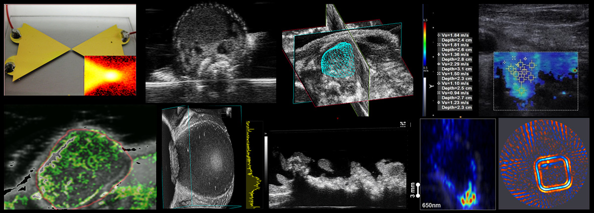
The Department of Medical Imaging is developing cutting-edge techniques and technologies to improve ultrasound contrast. For example, shear wave imaging to quantify elastic properties in human tendon, liver, and thyroid; hybrid modalities, such as photoacoustic, thermoacoustic, and acoustoelectric imaging to exploit interactions between ultrasound and other forms of energy (e.g., light, microwaves and electricity) to produce novel contrast for visualizing lymphatics, guiding cancer therapy, and functional brain imaging.
Other active project include the development of ultrasound contrast agents for the early detection of cancer or cardiovascular disease that can also be used as novel drug or gene delivery systems. Members of our team developed the vascular microbubble agent, Definity® , an FDA-approved echocardiographic contrast agent. Yet another project involves the development of new ultrasonic agents that deliver oxygen for reducing dose and improving outcomes for radiotherapies.


